Autofocussing, XYZ Sample scanning, Single-mode laser, water sample chamber,...
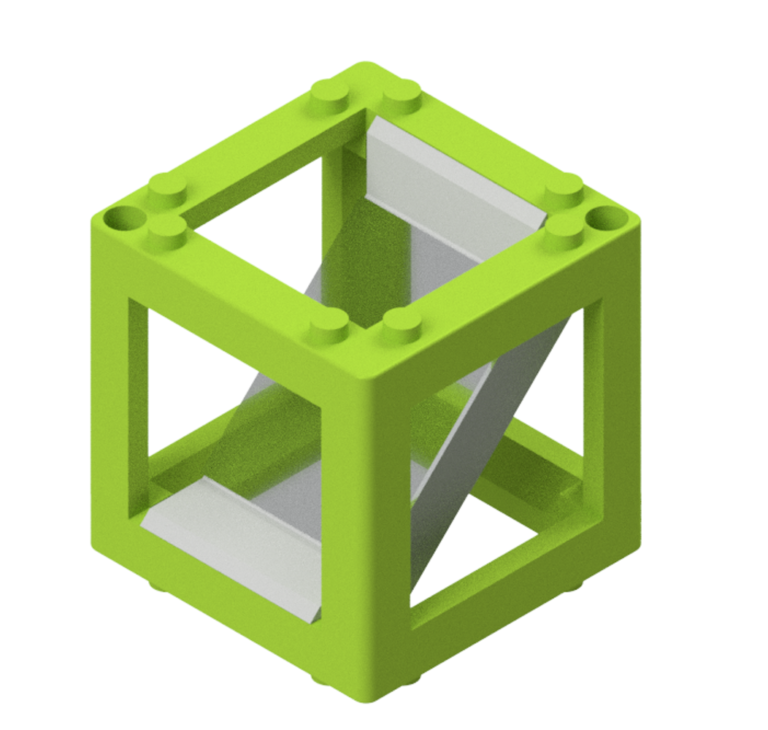
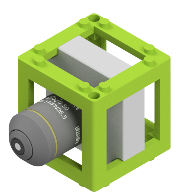
Modules
Enhance the functionality of your Scope
Community-driven Software
ImSwitch
Modular Microscopy Software
Open-Source
The adaptable System for your modular Idea
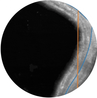
The resolution of any microscope is limited not only in the lateral direction but also in the axial direction. Light-sheet microscopy significantly enhances optical sectioning of a sample by using an illumination sheet perpendicular to the detection axis, selectively exciting the fluorescent sample. By moving the sample through the static light sheet and simultaneously capturing a stack of images, a 3D volume can be quickly generated. This method not only improves resolution along the optical axis but also offers the advantage of significantly reduced photodamage compared to confocal microscopy.
The openSPIM design has been around for years, often with a six-figure price tag, making it inaccessible to many. The openUC2 light-sheet benefits from the modular, cube-based arrangement of optical components, enabling rapid prototyping. This is especially advantageous for educational purposes, where one can learn the setup and alignment without the risk of damaging expensive components. Furthermore, this entry-level device is excellent for making custom hardware modifications by swapping modules or quickly testing new biological protocols (e.g., clearing). Intrigued? Reach out to us!
Completely autonomous it can work almost anywhere. With drivers even for Mac OS X and Jetson Nano/Raspberry pi, you can run the Light-Sheet almost anywhere
Light-Sheet Microscopy
Features
Objectives:
4x, NA=0.15, WD: 20mm, (FOV: 10mm, Res.: 2.3μm)
10x, NA=0.3 WD: 14mm, (FOV: 4mm, Res.: 1.2μm)
Illumination (Configurable):–
Transmission Brightfield LED
488nm Single-Mode, ca. 40 mW (Fluorescence)
635nm Single-Mode ca. 200 mW (Fluorescence)
488nm/635nm Single Mode Dual Colour Laser (Fluorescence)
Camera:
12 MP, USB 3.0, Sony IMX226
6 MP, USB 3.0, Sony IMX179
Objective Focusing Stage:
+/-25mm, 0.3nm increment open-loop
fully metall-based
Sample Stage:
+/-10mm, 0.3nm increment open-loop
fully metall-based
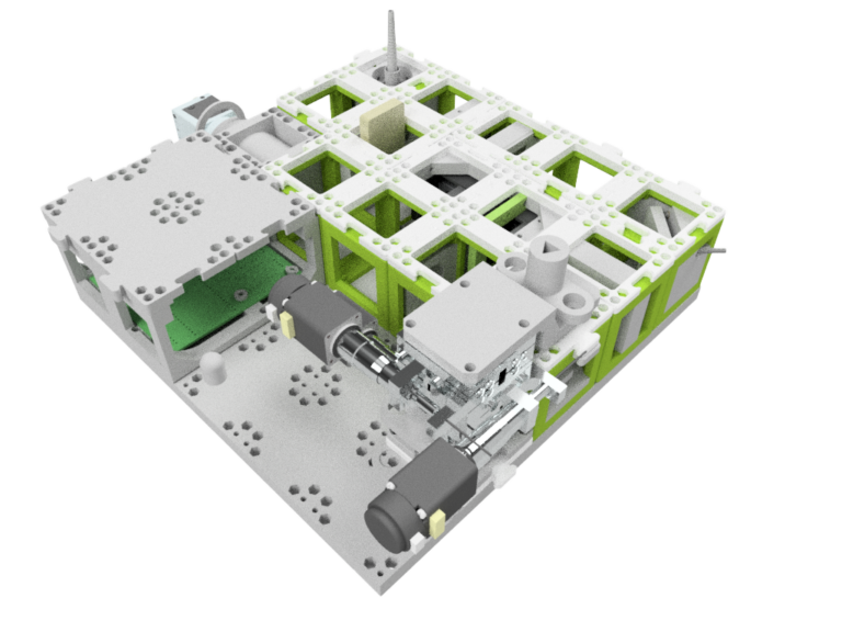
Sample Chamber:
3D printed, Volume ca. 10 x 10 x 10 mm^3
Two windows for excitation and emission
Inbuilt LED for transmission excitation
Magnetic quick-mounting mechanism
openUC2e:
ESP32-based
Laser, LED control
Stepper control
Powered by UC2-ESP firmware
Customizable
Adapt to your sample
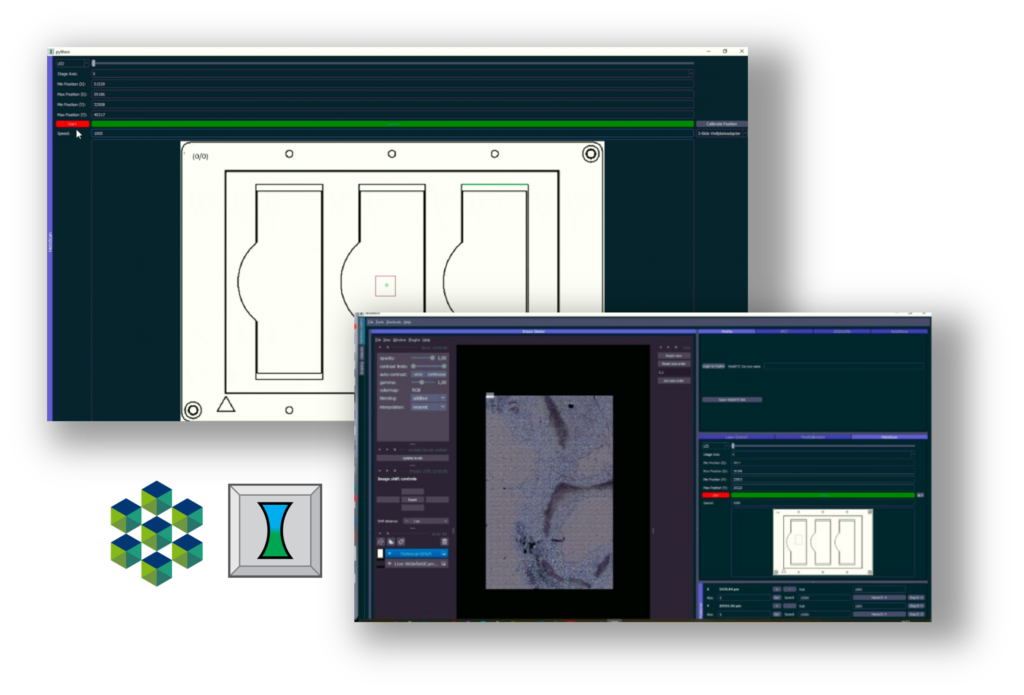
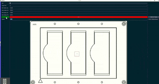
Discover the latest innovation in microscopy with our new XYZ stage, designed to enhance flexibility for both your application and samples. Effortlessly accommodate standard well-plates, or utilize our customized sample adaptors for seamless integration of any sample holder.
Our microscope sets itself apart by keeping the optics fixed, allowing the sample to move within a stationary focus, offering a unique approach compared to traditional designs. This open-source architecture not only facilitates easy customization but also simplifies the incorporation of additional components like cell incubation chambers.
The XY stage is powered by fastly moving stepper motors, achieving a resolution of ~0.3 µm, complemented by a magnetic encoder that ensures a control feedback loop as precise as 1µm. Moreover, the Z-axis benefits from a dual sub-micrometer precise linear translational stage mechanism, enabling smooth movement of the sample plate. Embrace unparalleled precision and adaptability with our innovative XYZ stage microscope, designed for advanced research needs.
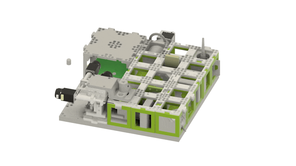
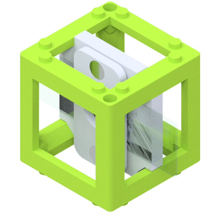
Customize any bit of the microscope by upgrading individual parts or complete subassemblies.
Modular
Customize your scope
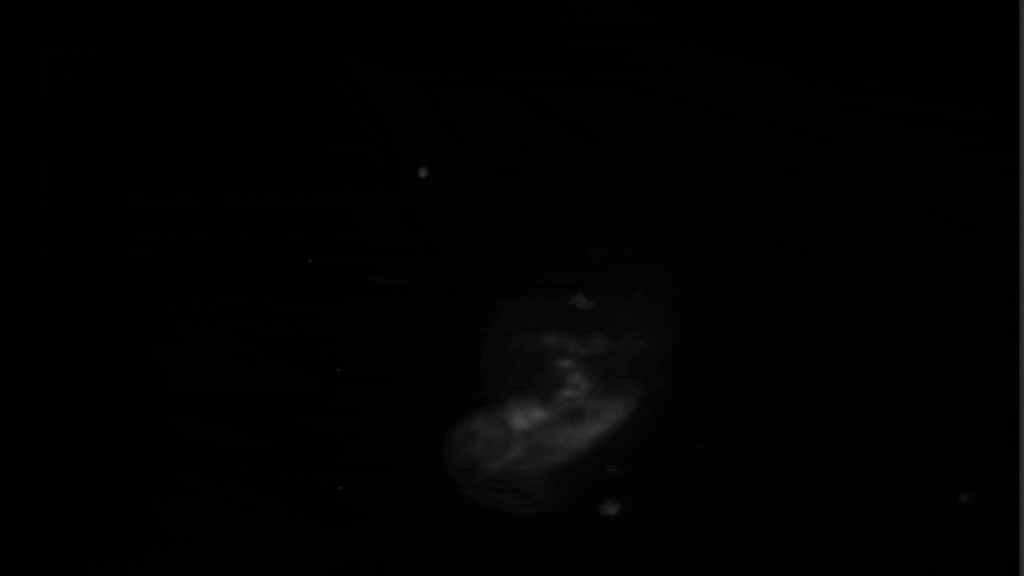
Quickly acquire 3D volumes such as the here visualized Zebrafish. Customize the sample holder to match your needs.
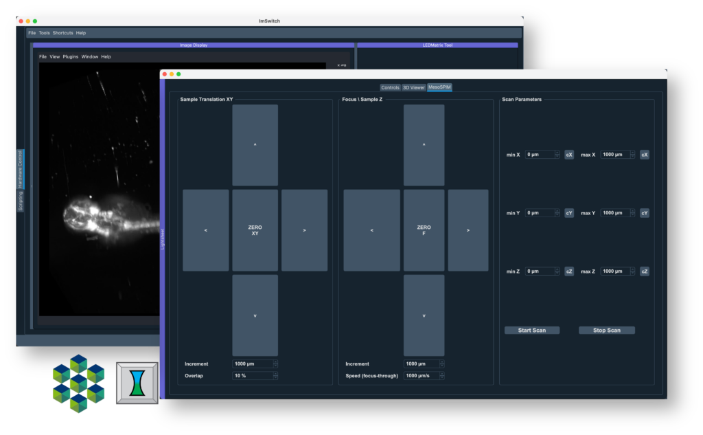
All interfaces of the device are fully open and can be controlled by the open-source microscopy control, acquisition, and processing software “ImSwitch.” This provides full control over all hardware and imaging components. Custom algorithms such as tracking, deconvolution, etc., can be integrated. The software features a Quickscan function that quickly captures and renders the volume during movement. Two different industry-grade CMOS cameras are available: one is compatible with both MAC and Windows, making it excellent for educational purposes, while the other, with significantly better noise statistics, is aimed primarily at research applications.
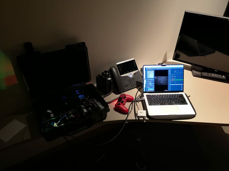
Portable
Field-trip ready.
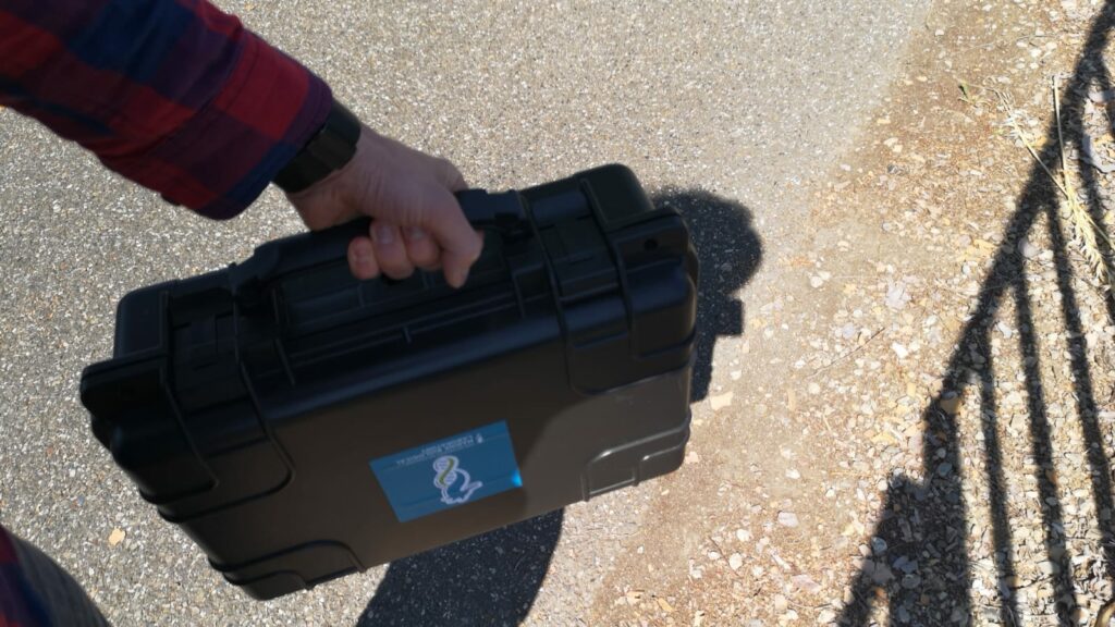
Bringing samples from field research back to your lab often presents immense challenges. Limited space or sample degradation can hinder the study of specimens, making on-site research highly advantageous. With the openUC2 system, this is no longer a problem. The robust case allows for transport to remote locations and even passes through airport security checks. Weighing just 3KG, it is not much heavier than a laptop. Powered by a 12V battery connected to your laptop, it offers maximum mobility and enables you to conduct experiments far from any power outlet.
The Light-Sheet is fully encapsulated and built inside a hard-shell case to survive field-trips or security checks at the airport.
The stage can load up to 4 slides at once and scans them in one go. ImSwitch offers multiple options to export the data for your workflow. Use it in Fiji, QuPath or your own libraries.
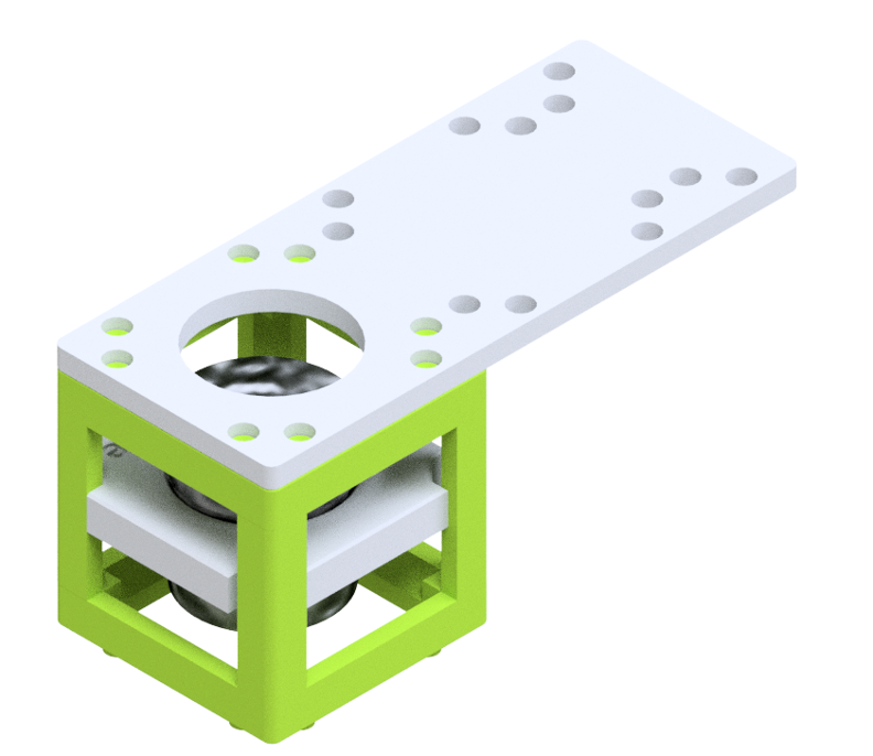
Mirror
2x mirrors (45°, front-surface). Change propagation direction of light to e.g. build compact setups
Lens
3x convex (2x 50mm, 1x 100mm), 1x concave (1x -50mm) lens Learn and understand basic ideas in imaging and combine them to build e.g. telescopes
RMS Mount
combination to image small samples and observe them e.g. by add
Sample Mount
Clip-based holder with which a specimen fixed on a carrier glass can be held in the beam path.